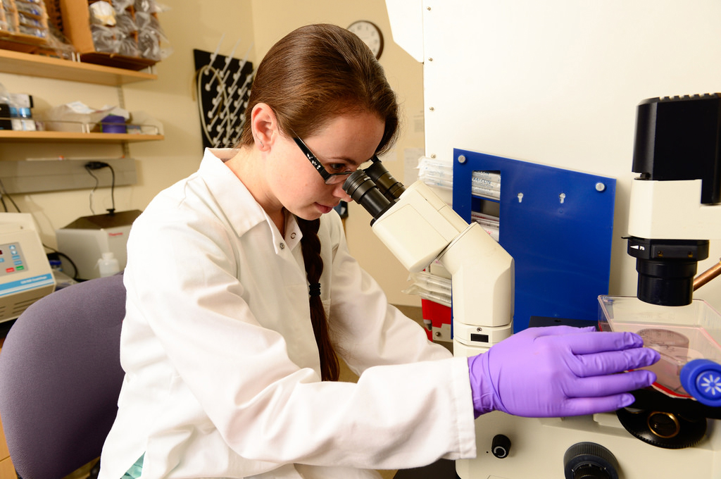
 Research Equipment Support
The CCAH launched its Faculty Equipment Grants Program in 2014 after recognizing the ongoing need of the school’s faculty to purchase new equipment and pay for replacements or repairs. “It’s hard to repair or replace equipment needed for research because most grants exclude equipment,” says Director Dr. Michael Kent. “If a freezer or centrifuge breaks, it needs to get done right away.”
In the program’s ten years of existence, over $2.9 million, has been used to finance over 225 total requests for equipment. During the 2025 round of funding, the CCAH fulfilled 22 requests — totaling more than $339,000 — for the following items:
Kowa Slit-Lamp SL-19 Plus
Principal Investigator: Bianca Da Costa Martins
The slit-lamp biomicroscope is essential for ocular exams because it provides the magnification needed to assess lesion depth. Photographing ocular lesions routinely requires patient sedation. Because the Kowa Slit-lamp SL-19 Plus has an integrated camera, it allows for real-time, high-magnification image acquisition during exams. Sedation is rarely required, which improves patient safety.
1.5mm Titanium Miniplate Kit
Principal Investigator: Boaz Arzi
The specialized Synthes 1.5mm titanium miniplate kit, used for mandibular and maxillofacial reconstruction in dogs, will be used for research and to train residents to perform reconstructive surgeries. The kit is clinically designated for complex maxillofacial fractures that require delicate fixation, particularly in challenging locations.
Surgical, Periodontics and Endodontics Instruments
Principal Investigator: Boaz Arzi
Dental and Oral Surgery Service residents will use these instruments to practice procedures on cadavers, enabling them to develop the high-level surgical skills needed for performing procedures on live patients.
Embedding Workstation
Principal Investigator: Clare Yellowley-Genetos
The Veterinary Orthopedic Research Laboratory (VORL) is focused on understanding the pathophysiology of multiple diseases associated with the musculoskeletal system, including arthritis, osteoporosis, bone fracture, cancers, bone metastasis, and tendinopathy. With this equipment, we will collect tissue samples, embed them in paraffin wax, and then take extremely fine sections. Using specialized stains, we will be able to examine the tissue structure and cellular biology of disease.
Pathology Microscope/Camera
Principal Investigator: Clinson Lui
Pathology microscope cameras capture high-resolution digital images from tissue slides used for research and diagnostics.
Main Bearing Rebuild for VORL’s Musculoskeletal Tissue Mechanical Testing Machine
Principal Investigator: Denis Marcellin-Little
The Veterinary Orthopedic Research Laboratory (VORL) aims to find the best method for providing animals with artificial joints when their own joints fail from arthritis or injury. Successful fracture treatment or joint replacement relies on testing the strength and function of bone plates, screws, and implants. This equipment simulates the load an animal’s body places on surgical parts or tissue, while measuring strength and stability. Research using this equipment will improve treatment function and quality.
Beckman Coulter Airfuge Tabletop Microultracentrifuge
Principal Investigator: Hugues Beaufrere
The Beckman Coulter Airfuge is a specialized bench-top ultracentrifuge that removes excess fat (lipemia) from blood samples, enhancing our ability to analyze them. High levels of lipemia can interfere with important lipoprotein tests performed by standard biochemical analyzer and lipoprotein electrophoresis. By clearing samples, the Airfuge ensures accurate test results and enables lipoprotein analysis through sequential separation by ultracentrifugation. Ultimately, this instrument will improve diagnostic precision, advance research (especially in exotic animal medicine), and enhance care.
Benchtop pH Meter
Principal Investigator: Jennifer Larsen
The Amino Acid Laboratory uses the pH meter to prepare biological samples from both research studies and clinical cases for analysis. We must adjust these samples—like urine, blood, and food—to maintain pH levels and accurately analyze various nutrients, including amino acids, vitamins, and other compounds. This analysis helps us evaluate individual veterinary patients and investigate the impact of different foods or medical treatments. This information guides treatment decisions and improves pet foods.
Repair of -80 Freezer
Principal Investigator: Jennifer Larsen
The Amino Acid Laboratory freezer stores samples, including blood, urine, tissue and food at super low temperatures. Low temperatures are critical for maintaining the stability of blood nutrients, which we analyze to assess animal health. We use the results to evaluate individual veterinary patients and perform research to investigate the impacts of different foods or medical treatments. This information guides treatment decisions and improves pet foods.
Densitometer
Principal Investigator: Krista Keller
Densitometers allow for measurement of optical density. In microbiology, densitometry is frequently used to quantify microbial suspensions. Quantifying microbes (bacteria or fungi) within a suspension is an important part of many experiments. For example, labs must quantify microbes to evaluate which antimicrobials best treat infections and which disinfectants best remove microbes from the environment. While counting bacteria or fungi manually under a microscope is time-consuming, the densitometer allows rapid, one-second quantification by measuring how much light solids (microbes) in block, otherwise known as optical density.
Sebia Minicap Flex Capillary Electrophoresis
Principal Investigator: Melanie Ammersbach
This equipment will allow the clinical laboratory us to perform in-house protein electrophoresis. Protein electrophoresis is used to detect certain types of cancers, such as lymphoma and multiple myeloma. It also expands the panel of tests available to detect and monitor diabetic patients. Performing these tests in-house will reduce the time it takes to get results.
A.R.C. FOX (Vet) Diode Laser 810nm
Principal Investigator: Munashe Chigerwe
The FOX laser produces laser energy that is absorbed by the target tissue, allowing it to selectively destroy neoplastic (cancerous) cells while minimizing damage to surrounding tissue. This makes it a promising option for managing skin cancers and a strong complement to surgical excision. In addition to destroying cancerous cells, it enhances wound healing and tissue regeneration, which is especially beneficial following tumor excision.
Centrifuge
Principal Investigator: Michael Kent
This laboratory centrifuge prepares samples from patients in radiation oncology clinical trials for flow cytometry. This process allows us to see the different types of immune cells in cancer patients.
CO2 Incubator, Plate Spinner, Manual and Electronic Pipettes, Cryo Storage and Cart
Principal Investigator: Robert Rebhun
The CO2 incubator is required to culture tumor cells and characterize their growth in the lab (in vitro), evaluate essential proteins and signaling pathways, and test drugs or therapies against these cells in the lab. The plate spinner, vortex mixer, pipettes, a laboratory cart, and storage boxes are essential to routine laboratory procedures for evaluating cancer cells in the lab and clinical cancer specimens.
VCCT Refrigerated Clinical Centrifuge and Label Printer
Principal Investigator: Robert Rebhun
A refrigerated benchtop centrifuge allows for the rapid and optimal processing of blood samples from patients in clinical trials. Analysis of these samples is critical for our trials testing novel drugs or immunotherapies as it helps determine levels of drug or antibodies in the blood. After processing, samples are stored in ultra-low temperature freezers or liquid nitrogen. Because normal labels freeze and fall off at these temperatures, a specialized label printer will ensure samples can be properly identified over time.
TissueLyser LT
Principal Investigator: Ryan Toedebusch
The TissueLyser LT effectively disrupts human and animal tissues as well as bacteria and yeast allowing reproducible yields of DNA, RNA, and protein in subsequent purification procedures. It even processes difficult-to-lyse tissues like heart and brain with the resulting DNA, RNA, and protein remaining intact. This capability enhances workflows and speeds up sample preparation for ongoing studies examining canine brain tumors, exploring neuroscience questions, and developing novel therapeutic avenues for canine diseases.
New Lens, Inspection and Warranty for Heidelberg Spectralis
Principal Investigator: Sara Thomasy
The Heidelberg Spectralis gives clinicians and clients unique views of ocular structures and improves patient care. By allowing clients to see ocular pathology in a new way, the device helps them make more informed treatment decisions. For example, when clients see how little corneal tissue is left with an infection using this technology, they often choose corneal surgery. The Spectralis also allows vision scientists to assess important eye structures to study their role in the disease process.
Professional Camera Equipment
Principal Investigator: Tyler Jordan
Veterinary dermatology relies heavily on visually assessing skin conditions for accurate diagnosis and effective treatment. Unfortunately, the field lacks standardized tools for evaluating and tracking skin lesions, making it challenging to measure treatment outcomes in clinical research. High-quality clinical images are essential for drawing evidence-based conclusions, comparing treatment options, and ensuring robust data for research publications. These professional-grade cameras would significantly enhance image quality and improve research reliability, increasing the likelihood of publication in high-impact journals.
Lift Tables
These tables will enable ophthalmology, dermatology, and primary care research teams to perform examinations and procedures on large-breed dogs at appropriate heights, ensuring safety and comfort for all.
